Spectral domain optical coherence tomography may have a role to play in evaluating retinal changes in eyes with sickle cell retinopathy.
 |
| (c) UIC |
Dr Jennifer I Lim, the Marion H. Schenk, Esq., Chair in Ophthalmology for Research of the Aging Eye, Professor of Ophthalmology & Director, Retina Service at the University of Illinois College of Medicine, presented about her work related to sickle cell retinopathy at a conference recently. Dr Lim opined that OCT can be a good tool to determine the severity of sickle cell retinopathy, even when patients are asymptomatic. The presence of retinal thinning does not mean the patient requires any intervention; it just signifies that the patient should be monitored regularly for anatomic changes and vision loss.
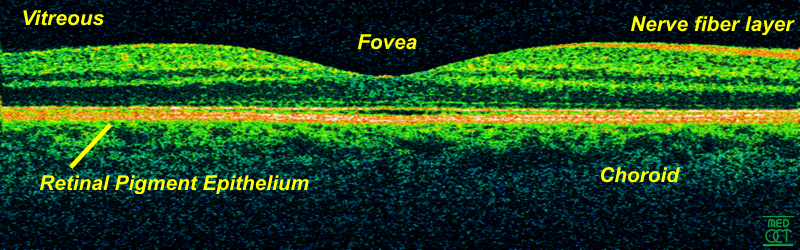 |
| OCT (c) Wikipedia |
In a study including 128 eyes of 64 sickle cell anemia patients and 24 eyes of 12 controls, Dr Lim reported that focal retinal thinning was observed in 36 sickle cell eyes (56%), while no control eyes showed it. Visual acuity in sickle cell eyes with focal macular thinning ranged from 20/15 to 20/200. Most eyes had 20/20 vision and were asymptomatic. Central macular thickness in sickle cell patients was 220 μm, while in controls, it was 240 μm.
In another part of the study, optic nerve and retinal nerve fiber layer thickness was measured in 151 eyes of 88 sickle cell patients and compared to 55 eyes of 30 controls. Results showed that sickle cell retinopathy eyes with macular thinning had thinner mean peripapillary RNFL in the nasal and superotemporal sectors than controls.
Choroidal thinning may also be associated with sickle cell disease.
Source
Read more on
Retina Global's website.
No comments:
Post a Comment
Thanks for your comments. We will get back to you shortly if there is a need to respond to it.
- Admin, Retina Global
Read more on Retina Global.