 |
| (c) Duke |
The research has been published online in the Journal of Neuroscience.
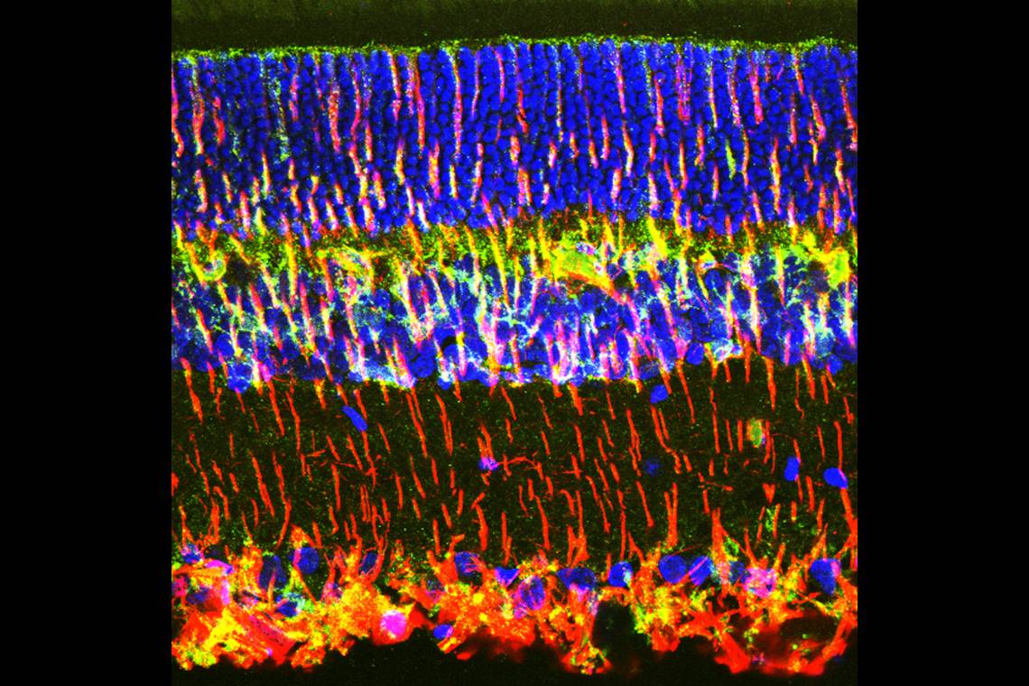
Retinas are built of a stack of different types of neurons, each connected by synapses that transmit signals from photoreceptors to the brain. Long, tree-shaped cells called Müller glia span the entire thickness of the retina, wrapping their branches around neurons to support their health and encourage the development of synapses.
Macular degeneration involves both the death of photoreceptor neurons -- the classic rods and cones that capture light and convert it into an electric signal -- and the loss of neural synapses within the retina.
Though age is the biggest risk factor for macular degeneration, genetics, race and lifestyle choices such as smoking also play a role.
The Duke scientists first examined the retinas of young rats that were genetically predisposed to an eye disease which causes progressive blindness similar to a disorder called retinitis pigmentosa in humans. They found that the neural synapses within the retina began to deteriorate even before the photoreceptors started to die.
As the number of neural synapses declined, the Müller glia also became sickly, pulling their branches away from neurons and dividing haphazardly.
When the researchers injected human umbilical stem cells behind the retinas of these rats, the Müller glia remained healthy, as did the neural synapses. The treatment succeeded in preserving the majority of the rats’ vision and stopped the photoreceptors from dying.
To test whether the Müller glia were truly the key players in the synaptic loss, the team used a gene-editing technique to remove a specific gene from Müller glia cells. Deleting this gene is known to cause retinal degeneration, but its function in Müller glia has never been explored.
Without this gene, the Müller glia were defective and bore striking similarities to those in rats that had developed retinitis pigmentosa. In addition, the neural connections within retinas of these rats were malformed, mimicking the problems seen in early stages of retinal degeneration.
This research indicates that Müller glia are important players in retinal health, as per the authors. They are impaired in disease, and effective cellular therapies should target not only other retinal cell types but these cells as well.
This research was supported by grants from the National Institutes of Health, the Duke University Chancellor’s Discovery Award, Kahn Neurotechnology Development grant and Regeneration Next Initiative Postdoctoral Fellowship. Parts of the work were performed under sponsored research agreements between Duke University and Janssen R&D.
Source
More about Retina Global here. We seek your support. Click here to donate.
No comments:
Post a Comment
Thanks for your comments. We will get back to you shortly if there is a need to respond to it.
- Admin, Retina Global
Read more on Retina Global.