Editor's notes:
To read more about what is Retinal Detachment, how is it caused, what are the symptoms, and what are the suggested things to do, click here.
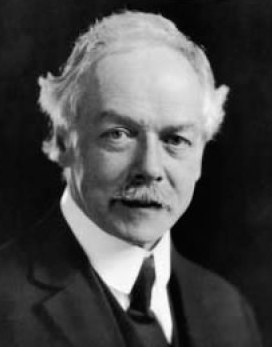
Retinal detachment was the first retinal pathology that could be managed with a surgical intervention. Dr Jules Gonin (pictured on the left - credit) came up with a solution to treat this condition. Before that, all patients with retinal detachment were doomed to blindness. His work from 1902 to 1921, when he recognized that a retinal break was the cause of a retinal detachment - and not a consequence as was largely believed those days - helped kickstart the specialty of retinal disease management. Though his technique, referred to as "ignipuncture", is now obsolete, his pioneering work laid the important foundation. Though his work did not gain recognition for quite some time, and was even opposed by many, he finally got his due in 1929 at the International Ophthalmology Congress in Amsterdam. Since then, his legacy has lived on in the eye hospital in Lausanne that bears his name, in the Gonin Medal awarded by the International Council of Ophthalmology, the Club Jules Gonin, and in a street named after his, the very street that he used to walk from home to the hospital every day.
Today, with management of retinal detachment that includes scleral buckle and vitrectomy, as many as ten percent of patients will ultimately suffer from permanent visual loss, usually due to the condition referred to as proliferative vitreoretinopathy (PVR).
Today, with management of retinal detachment that includes scleral buckle and vitrectomy, as many as ten percent of patients will ultimately suffer from permanent visual loss, usually due to the condition referred to as proliferative vitreoretinopathy (PVR).

The scientists & researchers have found more than 500 proteins that are produced following retinal detachment, many of which trigger the kind of inflammation that starts the scarring process. By identifying those proteins that are the most active, they can potentially begin developing experimental therapeutics that alter those proteins and hopefully either help promote tissue regeneration or inhibit scarring.
An estimated 57,000 people in the United States experience retinal detachment each year. The number is likely to be significantly more worldwide. The lifetime risk of developing a retinal detachment is about 1/300 and it remains a significant cause of legal blindness in the US, particularly if the macula has been damaged or if PVR develops, underscoring the need for better alternatives.
The scientists feel that the possible treatment options will not only help management of retinal detachment, but will also be helpful to people who have diseases like diabetes or macular degeneration. Having a non-surgical way to prevent retinal damage or help regenerate damaged tissue would impact millions of lives.
Dr. Cebulla’s identification of protein targets has been advanced significantly through the development of two novel animal models of retinal detachment. In order to identify the target proteins, she and her team developed a mouse model to conduct a proteomic study of how protein levels changed in response to retinal detachment. Using a technology called iTRAQ, the team was able to measure and monitor each individual protein to see how quantities changed over time, and then compare those to a control group.
With the target proteins identified, Dr. Cebulla then collaborated with neuroscientist Andy Fischer, PhD, at Ohio State’s College of Medicine, to develop the first ever chick model of retinal detachment. Chicks have a larger eye, making it easier for scientists to study the process of retinal detachment as well as the efficacy of experimental therapeutics versus smaller mammalian models. Unlike common mammalian models, chicks have good color vision, making their retinas rich in the type of cells found in a human eye.
Recently, Dr. Cebulla has started studying what happens inside the human eye after retinal detachment to supplement her observations from the animal models. During retinal detachment surgery, vitreous fluid is being collected from participating patients. Dr. Cebulla’s team will use the fluid sample, along with the patient’s blood to see how different proteins are being expressed.
Currently, the team has zeroed in on one protein in particular, and is testing a pharmacological agent that inhibits that protein to see if it can stop or slow the development of PVR and damage to the retina. They expect to have enough data to publish her next round of findings in just a few months.
Dr. Cebulla’s research is supported by The Ohio State Center for Clinical and Translational Science and the National Eye Institute.
Read about Retina Global here.
No comments:
Post a Comment
Thanks for your comments. We will get back to you shortly if there is a need to respond to it.
- Admin, Retina Global
Read more on Retina Global.