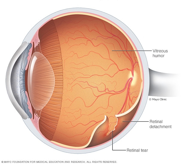
 |
| (c) Mayo Clinic |
Showing posts with label Retinal Detachment. Show all posts
Showing posts with label Retinal Detachment. Show all posts
Wednesday, November 28, 2018
Minimally invasive retinal detachment treatment offers patients better outcomes

Wednesday, April 15, 2015
Advancements in Retinal Detachment Research Pave the Road to Better Visual Recovery
Editor's notes:
To read more about what is Retinal Detachment, how is it caused, what are the symptoms, and what are the suggested things to do, click here.

Retinal detachment was the first retinal pathology that could be managed with a surgical intervention. Dr Jules Gonin (pictured on the left - credit) came up with a solution to treat this condition. Before that, all patients with retinal detachment were doomed to blindness. His work from 1902 to 1921, when he recognized that a retinal break was the cause of a retinal detachment - and not a consequence as was largely believed those days - helped kickstart the specialty of retinal disease management. Though his technique, referred to as "ignipuncture", is now obsolete, his pioneering work laid the important foundation. Though his work did not gain recognition for quite some time, and was even opposed by many, he finally got his due in 1929 at the International Ophthalmology Congress in Amsterdam. Since then, his legacy has lived on in the eye hospital in Lausanne that bears his name, in the Gonin Medal awarded by the International Council of Ophthalmology, the Club Jules Gonin, and in a street named after his, the very street that he used to walk from home to the hospital every day.
Thursday, July 24, 2014
Retinal detachment and its symptoms
What is a retinal detachment (RD)?
Retina detachment signifies, in a general sense, separation of the retina from its normal position.
What are the types of retinal detachment?
There are usually three types of retinal detachment, with the fourth being a combination of the first three.
- Rhegmatogenous retinal detachment (RRD) - the most common type that occurs due to a retinal or a break in the retina, allowing vitreous fluid to move into the sub retinal space and separate the retina from its normal position. These are usually managed by simple surgical interventions such as a scleral buckle (a band placed on the outside of the eye) or by vitrectomy.
- Tractional retinal detachment (TRD) - Usually seen in patients with diseases such as diabetic retinopathy, these usually occur due to forced pulling in the retina due to growth of fibrous tissue inside the eye. They do not have a hole in the retina. Management of these detachments usually requires vitrectomy to release of these 'tractional' forces.
- Secondary or serous retinal detachment - usually occurs, as the name suggests, due to some other reason inside or outside the eye. This can be from a benign growth in the eye, to cancer somewhere else in the body. Management includes treatment for the primary condition.
Subscribe to:
Posts (Atom)